Home
/ Animal Cell Microscope Slide - Watu Gwo Blog Animal Cell Light Microscope : Most cells, both animal and plant, range in size between 1 and 100 micrometers and are thus visible only with the aid of a microscope.
Animal Cell Microscope Slide - Watu Gwo Blog Animal Cell Light Microscope : Most cells, both animal and plant, range in size between 1 and 100 micrometers and are thus visible only with the aid of a microscope.
Animal Cell Microscope Slide - Watu Gwo Blog Animal Cell Light Microscope : Most cells, both animal and plant, range in size between 1 and 100 micrometers and are thus visible only with the aid of a microscope.. How to stain microscope slides. Mitosis of animal sec.(parascaris equorum ). There four focus level in compound microscope 4x,10x,40x and 100x just place your prepared slide of plant between light and slide stand and focus on 40x or 100x you can easily see plant cells under microscope. Add text 1) open the powerpoint slide in which you have to insert the text box. Onion cells onion cells under a microscope.
Preparing onion cell slides is a useful way to observe simple plant cells under the light microscope. Unlike animal cells (such as cheek cells) the cell wall of an onion and other plants are made up of cellulose, which protects the cell and maintains its shape. 9 cell microscopy (i) how did you apply the. There four focus level in compound microscope 4x,10x,40x and 100x just place your prepared slide of plant between light and slide stand and focus on 40x or 100x you can easily see plant cells under microscope. Are plant and animal cells the same?
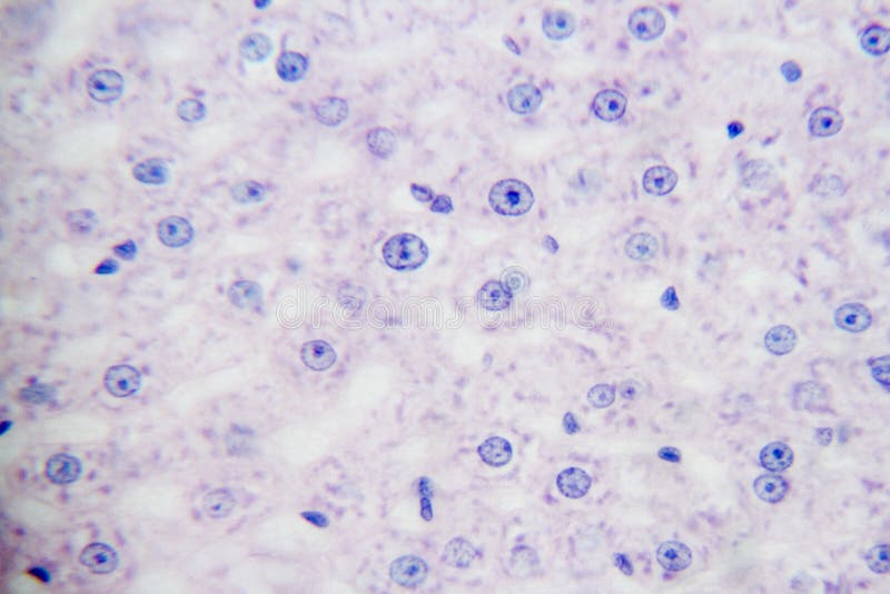
Improving Resolution Of Ordinary Light Microscope With Light Shrinking Material 2021 Wiley Analytical Science from analyticalscience.wiley.com Two slides demonstrating the cell membrane of an animal cell and the cell wall of a plant cell. Root tip of allium cepa l.s.(show mitotic division). Before cell division, the entire genome is copied. Squamous epithelium, isolated cells from human mouth. To prevent drying out or to protect lens or easier to view or keeps cells in place. Animal cell, section, microscope slide is a preparation of liver cells from the congo eel, amphiuma. Obtain cheek tissue by scratching. It prevents the slide from drying out when it's being.
Most cells, both animal and plant, range in size between 1 and 100 micrometers and are thus visible only with the aid of a microscope.
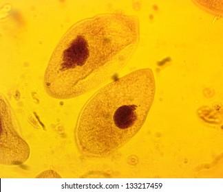
This is one of the tenets of the cell theory, a your microscope has four objectives of varying magnifications (4x, 10x, 40x, and 100x) mounted on a place a slide on the stage and use the mechanical stage controls to move it into place. This stain is often used to stain spores. Compact bone and hyaline cartilage t.s., two sections for comparison. This appears at the light microscope level at this stage in animal cells, there is a division and migration of the centrioles in the cytoplasm to initiate the formation of a spindle and asters. Onion cells onion cells under a microscope. Compound light microscope (lm) does not go beyond 2,000x magnificiation. All living things are composed of cells. Add text 1) open the powerpoint slide in which you have to insert the text box. Published on december 9, 2013 at 8:13pm by glenda stovall under cell. Animal and plant cells undergo a precise type of division called mitosis. Root tip of allium cepa l.s.(show mitotic division). Squamous epithelium, isolated cells from human mouth 2(d). A cell is a very tiny structure which exists in living bodies.
Cell structure is readily seen under low to medium power. It prevents the slide from drying out when it's being. Cleavage stage of egg of frog sec. Then click on the insert tab' in the ribbon and then inside the. 8 cell microscopy when preparing a sample of animal cells for examination under the microscope, after staining, a cover slip is placed on the slide.
Generalized Animal Cells Stock Image Image Of Anatomy 169339913 from thumbs.dreamstime.com This appears at the light microscope level at this stage in animal cells, there is a division and migration of the centrioles in the cytoplasm to initiate the formation of a spindle and asters. Cleavage stage of egg of frog sec. How to stain microscope slides. Squamous epithelium, isolated cells from human mouth 2(d). Typically the object is mounted (secured) on the slide, and then both are inserted together in the microscope for viewing. Mitosis of animal sec.(parascaris equorum ). Preparing onion cell slides is a useful way to observe simple plant cells under the light microscope. Add text 1) open the powerpoint slide in which you have to insert the text box.
A microscope slide is a thin flat piece of glass, typically 75 by 26 mm (3 by 1 inches) and about 1 mm thick, used to hold objects for examination under a microscope.
8 cell microscopy when preparing a sample of animal cells for examination under the microscope, after staining, a cover slip is placed on the slide. Published on jul 2, 2013. Preparation of cheek cell slide and viewing under a light microscope. Give a reason for this. Cell structure is readily seen under low to medium power. Animal and plant cells undergo a precise type of division called mitosis. Microscope cell staining is a technique used to enable better visualization of cells and cell parts under the microscope. How to stain microscope slides. 9 cell microscopy (i) how did you apply the. Before cell division, the entire genome is copied. Jiusion 48pcs prepared microscope slides specimen animals insects plants flowers sample biological specimen, stereo microscope slide for kids children students enlighten education. Typical animal cell pinocytotic vesicle lysosome golgi vesicles golgi vesicles rough er (endoplasmic reticulum) smooth er (no ribosomes) cell (plasma) 9. This appears at the light microscope level at this stage in animal cells, there is a division and migration of the centrioles in the cytoplasm to initiate the formation of a spindle and asters.
Compound light microscope (lm) does not go beyond 2,000x magnificiation. Unlike animal cells (such as cheek cells) the cell wall of an onion and other plants are made up of cellulose, which protects the cell and maintains its shape. Compact bone and hyaline cartilage t.s., two sections for comparison. How are varieties of living things organized? Epithelium cells of cavitas oris of humanw.m.
Animal Cell Under Microscope High Res Stock Images Shutterstock from image.shutterstock.com Animal and plant cells undergo a precise type of division called mitosis. Microscope cell staining is a technique used to enable better visualization of cells and cell parts under the microscope. Compact bone and hyaline cartilage t.s., two sections for comparison. Then click on the insert tab' in the ribbon and then inside the. 12 selected microscope slides of animal cytology. The thin membrane from between the layers of a raw preparing animal cell (cheek) wet mount. Obtain a slide and cover slip. Recommended for beginning microscopists or those studying the vertebrate zoology of amphibians.
Most cells, both animal and plant, range in size between 1 and 100 micrometers and are thus visible only with the aid of a microscope.
Animal cell, section, microscope slide is a preparation of liver cells from the congo eel, amphiuma. Squamous epithelium, isolated cells from human mouth 2(d). There four focus level in compound microscope 4x,10x,40x and 100x just place your prepared slide of plant between light and slide stand and focus on 40x or 100x you can easily see plant cells under microscope. Unlike animal cells (such as cheek cells) the cell wall of an onion and other plants are made up of cellulose, which protects the cell and maintains its shape. Published on december 9, 2013 at 8:13pm by glenda stovall under cell. Root tip of allium cepa l.s.(show mitotic division). Add text 1) open the powerpoint slide in which you have to insert the text box. Compound light microscope (lm) does not go beyond 2,000x magnificiation. Compact bone and hyaline cartilage t.s., two sections for comparison. Animal and plant cells undergo a precise type of division called mitosis. It prevents the slide from drying out when it's being. Plant and animal cells can be studied in greater detail with a. Then click on the insert tab' in the ribbon and then inside the.
Share :
Post a Comment
for "Animal Cell Microscope Slide - Watu Gwo Blog Animal Cell Light Microscope : Most cells, both animal and plant, range in size between 1 and 100 micrometers and are thus visible only with the aid of a microscope."

Post a Comment for "Animal Cell Microscope Slide - Watu Gwo Blog Animal Cell Light Microscope : Most cells, both animal and plant, range in size between 1 and 100 micrometers and are thus visible only with the aid of a microscope."